Eocene sepioid cephalopods - Belosaepia
This web page prepared by T. E. Yancey
Eocene age sediments along the northern Gulf of Mexico, especially in Mississippi
and Texas, contain some very well preserved specimens of sepioid cephalopods
(relatives of cuttlefish). These are often considered to be an oddity, because
they produced a skeleton with a tooth-like projection at the end that has
a minor resemblance to the jaw parts of a squid or octopus. Consequently the
fossils are often called "squid beaks" by collectors. However, the
internal part of the skeleton contains septa, showing that they are chambered
cephalopods. A typical example of the most common species of these fossils
is shown below.
Click on pictures to magnify
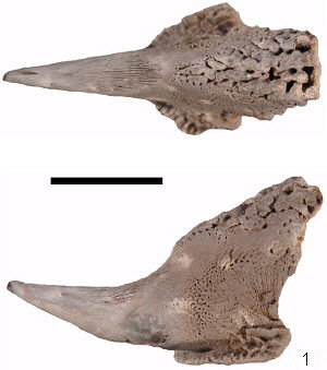
Figures 1,2: A nice specimen of Belosaepia ungula Gabb, 1860. The scale bar is 1 cm long.
The main parts of the belosaepiid skeleton are the
prong (the tooth-like part), the flat basal collar-like corona and the heavy
knobby callus on the top. The open space containing septa is rarely preserved
because it is weak and crushes easily when deposited in sediment. Usually
only the posterior end of the skeleton is preserved and more complete skeletons
are very rare.
The specimen shown above is better than average for Belosaepia, but
nearly all Belosaepia skeletons preserve only 5% to 10% of the skeleton
secreted by the animal and this one preserves only about 10%. The specimen
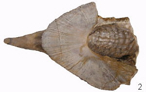
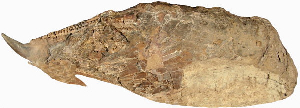
below (#3) is an unusually complete skeleton (about 90% complete), revealing
that the whole skeleton is much different then the normal fossil. This is
one of 4 nearly complete skeletons known for the genus. Most of the skeleton
is weak and breaks apart, leaving only the heavy, solid tooth-like part of
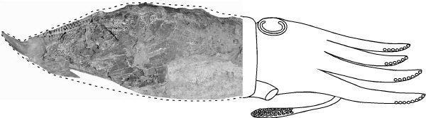
the skeleton as a recognizable fossil. A reconstruction of the living animal
(#4) shows the solid tooth-like prong is at the back (posterior) end to the
skeleton.
 3
3
 4
4
Figures 3,4: A nearly complete skeleton of B.
ungula from Brazos County, Texas and a reconstruction of the animal
with skeleton. The dashed line indicates that the outer surface of the body
tissue and shows that the skeleton is internal. Some of the lower margin is
reconstructed.
Although different in appearance, the belosaepiid fossil is most similar to
the cuttlebone of the common living cuttlefish, a cephalopod that is common
from Europe and Africa to east Asia. In contrast to the chambered condition
of a cuttlebone, Belosaepia has a large amount of solid skeleton, but
there are similar features in both Belosaepia and Sepia. The
figure below shows some comparable characters on each skeleton. Belosaepia
is an ancestor of the modern Sepia.
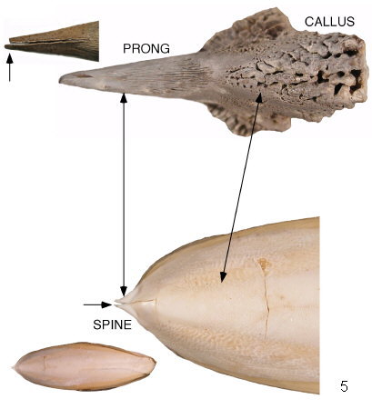
Figure 5: The prong of Belosaepia is equivalent to the spine of Sepia; the callus of Belosaepia is an over-developed equivalent of the rough upper surface of the cuttlebone. Both features are more developed on Belosaepia. The arrows point to an odd feature of sepioid skeletons: the prong/spine on these skeleton is easily split along a median plane. It is not known why this plane of weakness develops in the sepioid skeleton. It is apparently a surface with a higher content of non-mineral tissue.
Belosaepia skeletons were secreted as an internal
skeleton and were entirely covered with tissue; the animal secreting regular
layers one on top of the other. Like the rings in wood grown by a tree. One
of the surprising aspects of the belosaepiid skeleton is that late in life,
the animal started to dissolve much of the skeleton mass, leaving a reduced
and etched remnant of the well developed skeleton. Photos below (#6) show
an etched skeleton created by resorbing part of the skeleton mass. This occurred
late in life and presumably was related to reproduction. (Sepiids reproduce
only once, then die.) The occurrence of etching during life can be recognized
by looking at the sides of a skeleton. If the edges of growth layers in the
skeleton are visible (#7), it means that skeleton was dissolved by the animal.
An abraded surface or a broken surface looks different.
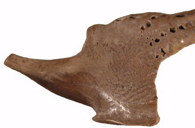 6
6 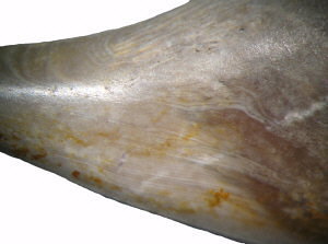 7
7
Figures 6,7: Appearance of belosaepiid skeletons resorbed late in life by the animal. Compare with the skeleton shown at top of the page. Resorbtion produces a smooth, shiny surface and thinning. A closeup view of the surface reveals the edges of growth lines, indicating truncation by dissolution.
A detailed study of Belosaepia ungula,
the most common belosaepid fossil in North America, was published in the 2010
March-April issue of the Journal of Paleontology. This report documents the
large and small features of the skeleton and some of the microstructure of
the skeleton. Belosaepiid fossils from the northern Gulf of Mexico region
are among the best preserved fossils of belosaepiids known. A companion study
of the sepioid genus Anomalosaepia is presented in the Journal of Paleontology,
vol. 85, p. 904-915 (see below).
The right to distribute digital copies of these articles
was arranged with the Paleontological Society.
[pdf]
Yancey, T.E., Garvie, C.L. and Wicksten, M., 2010, The Middle Eocene Belosaepia
ungula (Cephalopoda: Coleoida) from Texas: Structure, ontogeny and
function; Journal of Paleontology, v. 84, no. 2, p. 267-287.
[pdf]
Yancey, T.E., and Garvie, C.L., 2011, Redescription of Anomalosaepia
(Cephalopoda: Coleoida): a sepioid with a bimineralic calcite and aragonite
skeleton; Journal of Paleontology, v. 85, no. 5, p. 904-915.
Eocene sepioid cephalopods - Anomalosaepia: Click Here
![]()
Top of Page