Sponge spicules
Photography and sample preparation by Dr. Yuri
V. Yashunsky
Click on pictures to magnify
Sponge spicules
Photography and sample preparation by Dr. Yuri V. YashunskyClick on pictures to magnify
Discussion:
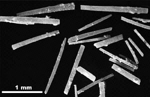
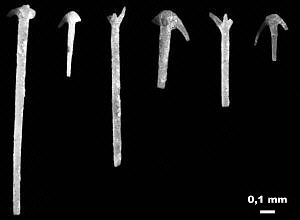
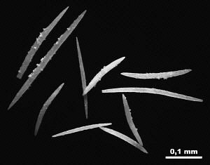
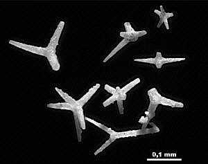
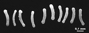
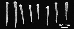
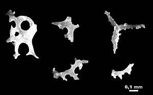
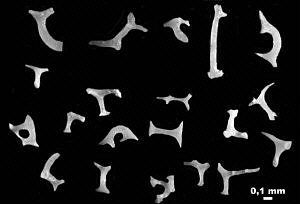
These photos show several types of siliceous sponge spicules that form
the support structure for the tissue of a sponge. All sponges have spicules
to support the pores and canals that carry water through the sponge. The
spicule support is needed to keep the canals open and allow it to feed
because the tissue is thin and weak. These photos show both the larger
spicules (megascleres) that provide major framework support and the
abundant small spicules (microscleres) that surround pores and provide
support to the wall in areas between the larger framework spicules.
Larger spicules that provide major framework support are usually fused
together. Although many spicules have the shape of a rod - straight or
curved - there are also many types of spicules that have multiple axes,
like those shown in several of the photos. The most common multi-axon
spicules are the triaxon type. Note the tiny nodes and spikes on the
sides of the spicules shown here.Many sponges secrete silica spicules, usually in combination with other
types of spicules. The most common sponges are demosponges that
secrete non-mineralized spongin spicules, but they also tend to secrete
some silica spicules as well. The spicules shown here are probably from
more than one species of sponge, although they might all occur in a single
species. Many different species of sponges secrete spicules of the same
shape. It is the arrangement of the framework spicules forming the skeleton
that provides a basis for recognition of species.
Go-Back
Phylum Porifera (sponges)
Fossil MenuSite Search Engine
search Carboniferous fossils of Russia or the Web