Fossil Images
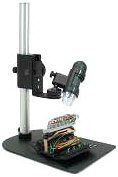
Captured with the Dino-Lite AM413T with
Rigid Stand
1.3 Megapixel, 1280x1024 resolution

Click on microscope for full description
Scanned images of these fossils are also showcased
on the
St. Louis Pennsylvanian Fossils of the Altamont Formation website
Fossil Menu
Gastropod - Pennsylvanian
Trachydomia nodosa (Meek & Worthen, 1861)
Magnification: 20x
Distance: 7 inches
LED lights useless at this distance
image captured using external lighting
Click
Here to Magnify
Click Here to view
Large Original image (20x)
Click
Here to view the web page with scans of this fossil
Back-Arrow to return to this page
Chiton (tail valve) - Pennsylvanian
Glaphurochiton carbonarius (Stevens, 1858)
Magnification: 20x range
Click
Here to Magnify
Click Here to view
Large Original image (20x)
Note the blue box
Click Here to Magnify
Click Here to view
Large Original image (200x)
Click Here
to view the web page with scans of this fossil
Back-Arrow to return to this page
Inarticulate Brachiopod - Pennsylvanian
Orbiculoidea missouriensis (Shumard, 1858)
Brachial Valve - Predator borings
Click
Here to Magnify
Click Here to view
Large Original image (20x)
Magnification: 200x
Click
Here to Magnify
Click Here to view
Large Original image (200x)
Click Here
to view the web page with scans of this fossil
Back-Arrow to return to this page
My first attempt imaging fossils
The rigid stand is not a Luxury - it's a Necessity. There are reasons why rigid stands are included when purchasing standard microscopes.
To bring an image into focus is no more complicated than using a standard microscope.
Resolution: Images were captured and saved at the highest resolution (1280x1024). These larger HQ images then can be cropped and re-sized (reducing the size of images improves the focus) saving the original image for future reference.
Additional lighting was required when imaging Trachydomia nodosa because increased distance required. Actually, I turned off the LED lights and used external lighting. Additional lighting not only illuminates the subject but also increases focus.
The scope's camera performed well capturing true colors.
The "micro-touch" button on the scope - I did not use "micro button" on the scope to take the pictures. Instead I clicked the mouse. Any movement of the scope can result in blurred images.
The software (including adding measurements to pictures) is straight forward and user friendly.
Focusing images on the computer monitor before capturing the pictures was a new and rewarding experience.
The hand-held scope (obviously) will not take the place of my camera with macro lens & ring light or the camera I attached to the microscope. However, the handheld scope definitely has its place on the the shelf with the other cameras.
Close-up focused imaging of fossils is a time consuming chore, even for experienced photographers. When I first saw a demonstration of the digital scope...Wow! The endless applications to our hobby hit me! One no longer needs to know a lot about photography in order to capture and share focused images. All can easily recognize a focused image on the computer monitor. When anyone can capture focused images to share with others...then that is "One Giant Leap" for Paleontology.
If you know how to use a standard microscope to bring
an image into focus (adjustments in distance, magnification, and lighting)
then you are ready for a visual treat capturing images without leaving the
comfort of your computer chair. When the image is in focus...Click
Barry Sutton
Top of Page
Go-Back
Digital handheld microscopes
Fossil Images: Copyright 2010 © LakeNeosho.org
- All Rights Reserved
Contact: Barry Sutton
BGSutton@LakeNeosho.org